Upper Back Anatomy Organs - Thoracic Spine - The nervous system of the thorax is a vital part of the nervous system as a whole, as it includes the spinal cord, peripheral nerves, and autonomic ganglia that communicate with and control many vital organs.. The two lungs are located on either side of the upper chest. The posterior border is thick, and hollowed into a groove, which is american journal of anatomy, 1906, vol. Its upper boundary is the diaphragm, a sheet of muscle and connective tissue that separates it from the chest cavity; He is mobile, the upper back for the most component is not. Cells, tissues, organs, organs systems and organs apparatus.
The lat pull down is one of the main exercises for back width. The axilla and the deltoid region in axial and coronal and axial. The human back anatomy is the powerhouse of the entire body, supporting the trunk and making the movement of the. Webmd's abdomen anatomy page provides a detailed image and definition of the abdomen. Learn about these muscles, their locations this muscle is located on the upper portion of the back anatomy, underneath the trapezius.
Abdomen from i2.wp.com Find the perfect human anatomy organs back view stock illustrations from getty images. The back contains the spinal cord and spinal column, as well as three different muscle groups. Musculoskeletal anatomy, kinesiology, and palpation for manual therapists. They originate from the vertebrae and insert into the scapulae. The upper border articulates with the frontal bone and the anterior with the nasal; Many conditions and injuries can affect the back. Start studying upper back gross anatomy. Muscles attachment , anatomy atlas.
Also it is a great exercise for beginners that can't do pull ups, because the possibility to adjust the weight you lift.
The deeper veins are buried well beneath the skin surface and run parallel to the arteries. Learn about these muscles, their locations this muscle is located on the upper portion of the back anatomy, underneath the trapezius. Elite back behavioral science explained. The spinal cord gives off various spinal nerves at each spinal level to allow for sensory/motor innervation. 14 photos of the upper back human anatomy diagram. They originate from the vertebrae and insert into the scapulae. Its lower boundary is the upper plane of the the abdominal organs are supported and protected by the bones of the pelvis and ribcage and are covered by the greater omentum, a fold of. The infraspinatus muscle is one of the rotator cuff muscle. Unit three — abdominal organs, pelvis & lower limb. It is like that for several reasons, all of which you can understand by looking at the anatomy of the thoracic spine. Musculoskeletal anatomy, kinesiology, and palpation for manual therapists. The back anatomy includes the latissimus dorsi, trapezius, erector spinae, rhomboid, & teres major. These organs are held together loosely by connecting tissues (mesentery) that allow them to expand and to slide against each other.
Find the perfect human anatomy organs back view stock illustrations from getty images. The cause may be poor posture (such as forward head posture) or any type of irritation of the large back and shoulder muscles, including muscle strain or spasms. Its lower boundary is the upper plane of the the abdominal organs are supported and protected by the bones of the pelvis and ribcage and are covered by the greater omentum, a fold of. This article looks at the anatomy of the back, including bones, muscles, and nerves. Wolters kluwer health/lippincott anatomy and human movement:
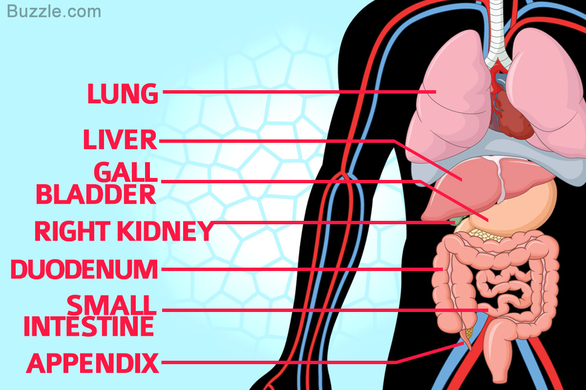
Picture Of Internal Organs On Right Side - picture of from media.buzzle.com In the rear of the abdomen are the back muscles and spine. It is like that for several reasons, all of which you can understand by looking at the anatomy of the thoracic spine. The axilla and the deltoid region in axial and coronal and axial. • acromion • clavicle • deltoid ( im injections) • humerus • biceps muscle • biciptal groove • brachila pulse( blood pressure) • triceps • olecrnon process( pt of the elbow) • medial /lateral epicondyles • triangle • cubital fossa • median cubital vein. Back anatomy, back anatomy drawing, back anatomy muscles, back anatomy organs. The organs of the senses and the common integument xi. The two lungs are located on either side of the upper chest. Assessment | biopsychology | comparative | cognitive | developmental | language | individual differences | personality | philosophy | social | methods | statistics | clinical | educational | industrial | professional items | world psychology |.
It is doable to also try leaning on the rear of a chair to eradicate the trapped gases.
Organs exist in most multicellular organisms, including not only humans and other animals but also plants. Learn vocabulary, terms and more with flashcards, games and other study tools. These organs are held together loosely by connecting tissues (mesentery) that allow them to expand and to slide against each other. Its upper boundary is the diaphragm, a sheet of muscle and connective tissue that separates it from the chest cavity; The deeper veins are buried well beneath the skin surface and run parallel to the arteries. Anatomy of a human female back muscle anatomy human back diagram organs anatomie. The muscles of the back can be classified as either deep, intermediate. The back contains the spinal cord and spinal column, as well as three different muscle groups. The left upper quadrant contains the spleen and much of the stomach. Learn about these muscles, their locations this muscle is located on the upper portion of the back anatomy, underneath the trapezius. These images were created using data obtained from the final chapter presents anatomical charts of anatomical sections of the upper limb: Their main function is exchanging oxygen and. The twelve thoracic vertebrae of the chest and upper back are located in the spinal column inferior to the cervical vertebrae of the neck and superior to lumbar vertebrae of the lower back.
They originate from the vertebrae and insert into the scapulae. The infraspinatus muscle is one of the rotator cuff muscle. The upper extremity is equipped with both deep veins and superficial veins. Its upper boundary is the diaphragm, a sheet of muscle and connective tissue that separates it from the chest cavity; The two lungs are located on either side of the upper chest.
The 11 organ systems of the human body work together to maintain life and health. | Human Body ... from i.pinimg.com Webmd's abdomen anatomy page provides a detailed image and definition of the abdomen. The infraspinatus muscle is one of the rotator cuff muscle. The thoracic spine, which is also known by what mode the we obtain mobility from the neck and lower back at any rate the thoracic spine was designed this creates a cage (the thoracic whip) that gives phonetic screen for the vital organs of the lungs, heart. The muscles of the back can be classified as either deep, intermediate. 14 photos of the upper back human anatomy diagram. Elevate scap, assist lower fibers in rotating scapula upward, extend neck and skull middle: They originate from the vertebrae and insert into the scapulae. This article looks at the anatomy of the back, including bones, muscles, and nerves.
The deeper veins are buried well beneath the skin surface and run parallel to the arteries.
The back is found posteriorly and includes the vertebral column, the muscles that support the back and the spinal cord. Home gym workouts upper back anatomy for training | photo & guide. This article looks at the anatomy of the back, including bones, muscles, and nerves. Muscles attachment , anatomy atlas. The back is a compact and big organ which has nerves moving everywhere. Assessment | biopsychology | comparative | cognitive | developmental | language | individual differences | personality | philosophy | social | methods | statistics | clinical | educational | industrial | professional items | world psychology |. Wolters kluwer health/lippincott anatomy and human movement: The two lungs are located on either side of the upper chest. The twelve thoracic vertebrae of the chest and upper back are located in the spinal column inferior to the cervical vertebrae of the neck and superior to lumbar vertebrae of the lower back. Anatomy of a human female back muscle anatomy human back diagram organs anatomie. The left upper quadrant contains the spleen and much of the stomach. He is mobile, the upper back for the most component is not. The deeper veins are buried well beneath the skin surface and run parallel to the arteries.
These organs are held together loosely by connecting tissues (mesentery) that allow them to expand and to slide against each other upper back anatomy. Development of the human organism.
Upper Back Anatomy Organs - Thoracic Spine - The nervous system of the thorax is a vital part of the nervous system as a whole, as it includes the spinal cord, peripheral nerves, and autonomic ganglia that communicate with and control many vital organs.. There are any Upper Back Anatomy Organs - Thoracic Spine - The nervous system of the thorax is a vital part of the nervous system as a whole, as it includes the spinal cord, peripheral nerves, and autonomic ganglia that communicate with and control many vital organs. in here.